

Description:
The muscular leg model illustrates both the superficial and deeper muscles, eight of which are removable. Tendons, vessels, nerves and bone components of the left leg and foot are shown in great detail in the muscular leg. All parts of muscular leg numbered.


Description:
This life size functional hip joint model clearly shows the anatomy and mechanics of the human hip joint. Demonstrates abduction, anteversion, retroversion and internal/external rotation. This functional joint consists of portion of femur, hip bone and joint ligaments.

Description:
This life-size functional knee joint model clearly shows the anatomy and mechanics of the knee joint. Demonstrates abduction, anteversion, retroversion and internal/external rotation. Model consists of portion of femur, tibia and portion of fibula; also includes meniscus, patella with quadriceps tendon and joint ligaments, including the ACL and PCL.


Description:
All the superficial musculature of the human form is accurately reproduced and detailed in life-like colors in this desktop size of the muscular figure. The chest plate is removable from the muscular figure to reveal the internal organs and the right side contains a female mammary gland. Over 125 hand-numbered and identified structures of the human anatomy on this muscular figure.


Desription:
This life-size functional shoulder joint model shows the anatomy and mechanics of the shoulder joint. Consisting of the scapula, clavical, portion of humerus and joint ligaments, this fully flexible shoulder joint model clearly demonstrates abduction, anteversion, retroversion and internal/external rotation.

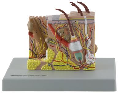
Description:
This model is composed of two parts and shows the structure of the human skin in three dimensions. Anatomical structures are shown in detail, including the hair follicles, sebaceous glands, erector muscles, Pacinian corpuscles, nerves and blood vessels. The different layers of the skin are clearly defined. A portion of a hair shaft can be removed to show internal details. Includes numbered key. Size (without base), 24 × 25 × 11 cm.


